The Brain's Faintest Signal: Inside the Cold, Silent Rooms Where MEG Makes Sense
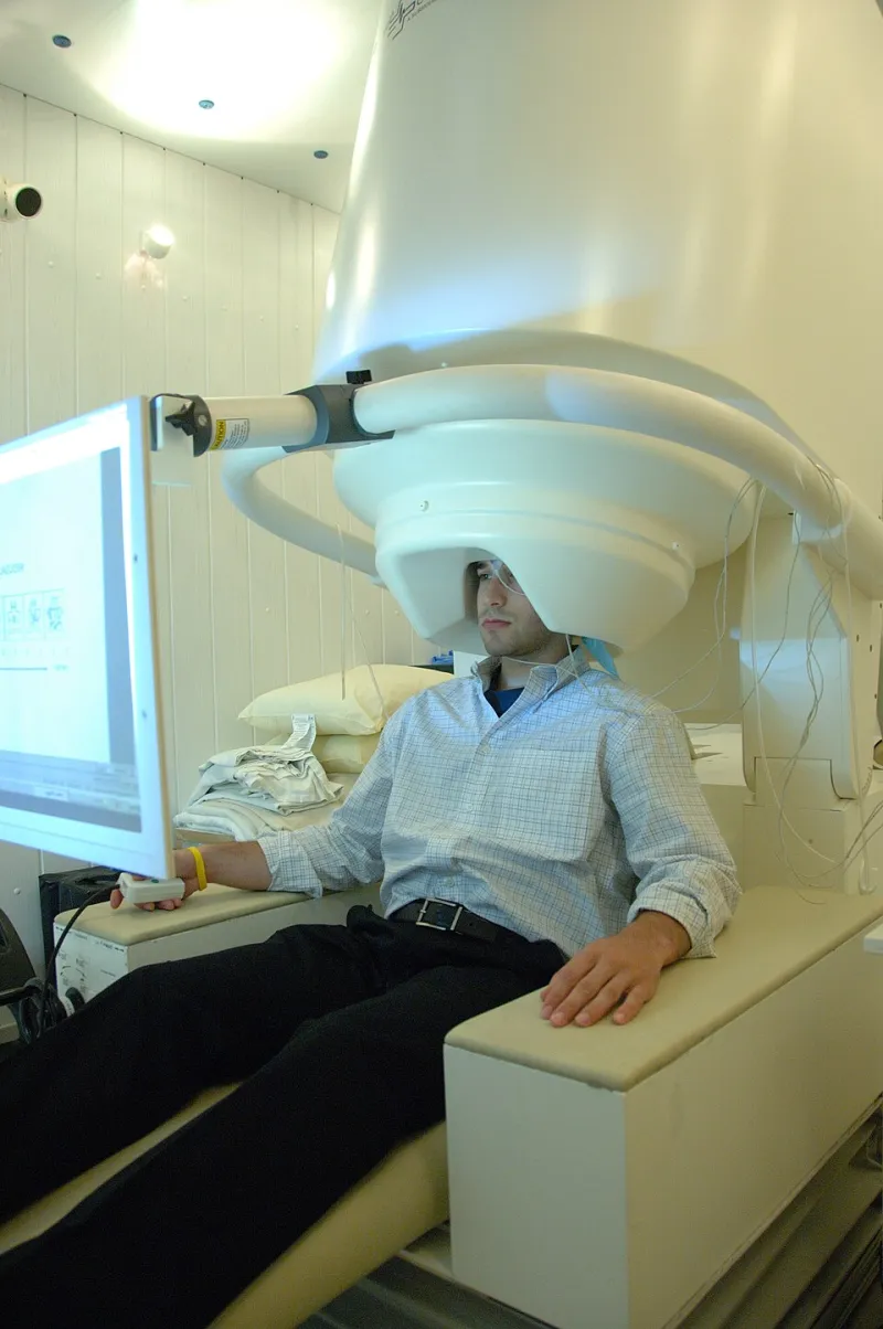
Photo: Unknown NIMH author, Public domain, via Wikimedia Commons
In a quiet corner of Cardiff, behind a door that looks more like a bank vault than a hospital entrance, someone is thinking about a word. They are not moving. They are not speaking. And yet, a few hundred sensors suspended around their head are picking up the magnetic signature of that thought — a field so faint it makes the Earth's own magnetism look like a foghorn.
This is magnetoencephalography, or MEG. It does not look like a scanner. There is no clanging, no tunnel, no radioactive tracer. Instead, there is a helmet, a very cold container of liquid helium, and a room designed to keep the rest of the world's magnetism out. The result is one of the most direct views we have of the brain at work, millisecond by millisecond.
A signal smaller than small
Every time neurons fire together, tiny electrical currents flow along their membranes. Electric currents make magnetic fields — that is not a quirk of biology, it is just physics. The trouble is scale. A decent MEG signal from the human brain sits somewhere around 10 to 100 femtoteslas. A femtotesla is a millionth of a billionth of a tesla. The Earth's magnetic field, which turns a compass needle, is about 50 microteslas. That means the brain's field is roughly a million times weaker than the field you are sitting in right now.
So how do you measure something that is overwhelmed by the magnetic noise of a passing tram, a lift motor, or the metal in your own fillings? You build a room that is almost magnetically dead.
The vault that keeps the world out
A MEG suite is usually wrapped in multiple layers of mu-metal, a nickel-iron alloy with a very high magnetic permeability. Think of it as a magnetic shortcut: field lines prefer to travel through the mu-metal rather than the air inside the room, so they get diverted around the patient. The layers are often demagnetised regularly, and the whole room is also shielded against radio-frequency interference. Even the lights and the air conditioning get chosen with care. In some labs, a passing lorry outside can still show up as a wobble on the traces.
The point is not luxury. It is silence. Without that shielding, the brain's magnetic whisper would be buried under the electromagnetic clatter of modern life.
Superconducting ears
The sensors themselves are another piece of quiet engineering. Most MEG systems rely on SQUIDs — superconducting quantum interference devices. A SQUID is a loop of superconducting material, usually niobium, interrupted by one or two Josephson junctions. Cool it below its critical temperature, and it becomes exquisitely sensitive to tiny changes in magnetic flux. A changing field induces a current, and the electronics turn that into a voltage you can record.
That sensitivity comes at a cost: the sensors have to sit at around 4 kelvin, just a few degrees above absolute zero. Liquid helium does the heavy lifting, which is why MEG labs have a slightly industrial feel — cryostats, dewars, and careful conversations about helium deliveries. A typical helmet-style array packs a few hundred SQUID sensors around the head, each one listening to a different patch of cortex.
The payoff is speed. Functional MRI, the workhorse of brain imaging, measures blood oxygenation and takes seconds. MEG measures the magnetic fields of neural currents directly, so it can resolve events on the millisecond timescale — fast enough to follow the choreography of a seizure, or the moment a sound registers in the auditory cortex.
What it is actually good for
In Britain, MEG is still largely a research tool. As of 2026, only a handful of systems operate across the country, mostly in university centres such as Cardiff University's Brain Research Imaging Centre, UCL, and a small number of other specialist sites. The NHS does not routinely offer MEG scans. If you are referred for a brain scan, the odds are overwhelming that you will get an MRI or a CT, not a MEG.
That is not because MEG is useless. Its clearest clinical niche is epilepsy. Before surgery for drug-resistant epilepsy, clinicians need to know where seizures start. MEG can localise that source with good spatial resolution and superb temporal resolution, sometimes catching activity that scalp EEG misses. It is also used in research on language, memory, autism, and brain development. Because it involves no ionising radiation and no loud noise, it can be gentler for children and for people who find MRI difficult.
But MEG has practical limits. It costs millions to buy and install, needs a shielded room and a helium supply, and demands specialist physicists and radiographers to run it. A scan is short, but the setup is not. Throughput is low. And the spatial resolution, while decent, is not as sharp as fMRI or invasive electrodes. It is a complement, not a replacement.
The wearable turn
The most interesting shift is towards sensors that do not need a fixed helmet. Optically pumped magnetometers, or OPMs, use lasers and caesium or rubidium vapour to measure magnetic fields. They can operate at room temperature, which means no liquid helium and no rigid cryostat. Wearable OPM helmets let people move their heads slightly during a scan, which matters for children and for anyone who finds stillness hard.
UK groups are part of that push, working alongside international teams to turn laboratory demonstrations into something more routine. It is early days. OPMs have their own noise problems, and a wearable helmet is only as good as its shielding. But the direction of travel is clear: smaller, cheaper, more flexible sensors that could one day bring MEG closer to ordinary clinical use.
For now, the quiet rooms remain rare. A MEG scan is still a small piece of physics theatre — cold sensors, silent walls, and a person asked to think about a word while a millionth of a whisper is coaxed into a graph. The magnet does not read your mind. It just listens to the electricity your neurons cannot help making.